Interviewer & Japanese Writer: Yamamoto Takaya; Translation & Editing: Matthew Cherry
Dr. Shinji Fuke was selected for the 2018 INNO-vation Program’s Disruptive Challenge for his research on detecting Congenital Heart Disease (CHD) before birth.
His theory was centered around taking sliced images of a healthy child’s heart using ultrasonic waves and layering them on top of each other, much in the same way a 3D printer works. By performing this task at three different angles, a 3DCG model of a healthy child’s heart can be created. Sliced images from the 3D model can then be layered on top of images of a child’s actual heart, allowing for easier detection of any irregularities.
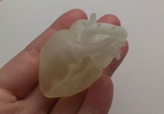
This model of a child’s heart was 3D printed using sliced images.
By progressing with this research, Dr. Fuke learned that he could draw conclusions about irregularities in the heart by using this method. He also discovered he could patent the method, but there was still a problem left to solve. “Ultrasonography and CT image data used in medical care must adhere to DICOM, the international standard protocol for such images,” Dr. Fuke explained. “DICOM files are a type of black box, meaning that they contain information that cannot be modified or altered in any way, so it’s not possible to remove images from them and lay them on top of other images.” Although he had shown his theory on detecting CHD before birth to be viable, it wasn’t possible to actually implement it. This is where his first challenge in 2018 came to a close.
It wasn’t until 2021 that he thought up a way to solve this problem. With his new idea in mind, he applied for the INNO-vation Program and was selected once again.
Software known as Materialise Mimics was the breakthrough for his research. This medical image processing software can read and load externally-created 3DCG model data. In 2018, the idea of laying 3DCG model data onto sliced images of a child’s heart was theoretically possible but infeasible to put into practice. Thanks to this software the process became a reality and would help make detecting irregularities in children’s hearts faster and easier.
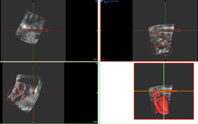
A 3DCG model is laid on top of images of a heart using Material Mimics.
This method still presented a hurdle to clear. Depending on the term of pregnancy, a child’s developing heart will differ in size and present individual characteristic differences. If it’s not possible to edit and alter the 3DCG model data to fit the images of a particular child’s heart, diagnosing irregularities through the image-layering process becomes impossible. Dr. Fuke knew that he could align the 3DCG model data with 7 distinct points in a fetus’s developing heart (such as the inferior vena cava and the descending aorta) by altering the model itself to fit the imagery. Special software would be required to make these alterations, but Dr. Fuke didn’t have any software development experience.
“I spoke with a lot of people about it, but most of them told me that even if I did receive funding for the software development, CHD only affects around 1 in 100 children, so it would never become a business,” Dr. Fuke explained. “It would be nice if a company or research lab could have helped me with the software development, but at that point, I figured there was no choice but to learn how to develop it myself.”
In Part 3, we’ll take a look at what Dr. Fuke has been doing since his time in the INNO-vation Program.